


Now Reading: Scientists Unveil First-Ever Complete Map of a Mammal’s Peripheral Nervous System
-
01
Scientists Unveil First-Ever Complete Map of a Mammal’s Peripheral Nervous System
Scientists Unveil First-Ever Complete Map of a Mammal’s Peripheral Nervous System
Fast Summary
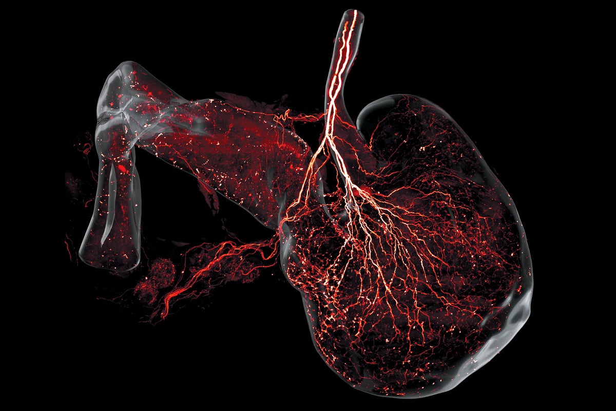
- News Title: “These Stunning Images Show Every Nerve in a Mouse.”
- Content: Researchers have produced the first-ever three-dimensional full-body map of a mammalian peripheral nervous system (PNS) using mice.
- study Details:
– Published in Cell on July 10, 2025, by Mei-Yu Shi et al.- Utilized techniques including immunostaining, genetic modifications for fluorescent neurons, and viral tracing of nerve projections.
– The mapping process involved making mouse bodies visually transparent and imaging their structure layer-by-layer at subcellular resolution, taking about 40 hours per mouse.
- Findings:
– Each vagus nerve fiber connects to only one organ in the gut rather than branching out to multiple organs as hypothesized earlier.
– Sympathetic nerves and cranial nerve patterns were also highlighted with precise details for various organs like kidneys and intestines.
- Meaning: Provides substantial groundwork for studying how the PNS regulates body functions and implications for precision surgeries or therapies targeting neurological disorders such as chronic pain.
Indian Opinion Analysis
This groundbreaking visualization of an animal’s entire PNS marks a significant advancement in neuroscience. For India-a nation increasingly focused on scientific research-it demonstrates the potential applications of cutting-edge technologies like high-resolution imaging combined with genetic engineering.If applied to human tissues next, this mapping may influence global medical practices in neurology and surgery. Institutions like those working on neuroscience within India could find inspiration from these methodologies to enhance local capacity-building initiatives aimed at treating nerve-related disorders prevalent among Indian communities.
Moreover, India’s investment focus on biosciences through organizations such as DBT (Department of Biotechnology) could be well-positioned to adapt similar innovations toward therapeutic advancements. Collaboration opportunities with international researchers may emerge that further India’s contributions while fostering advances in precision medicine globally.Read More


Related Posts
Stay Informed With the Latest & Most Important News
Previous Post
Next Post


Advertisement